English
 Հայերեն
 Русский
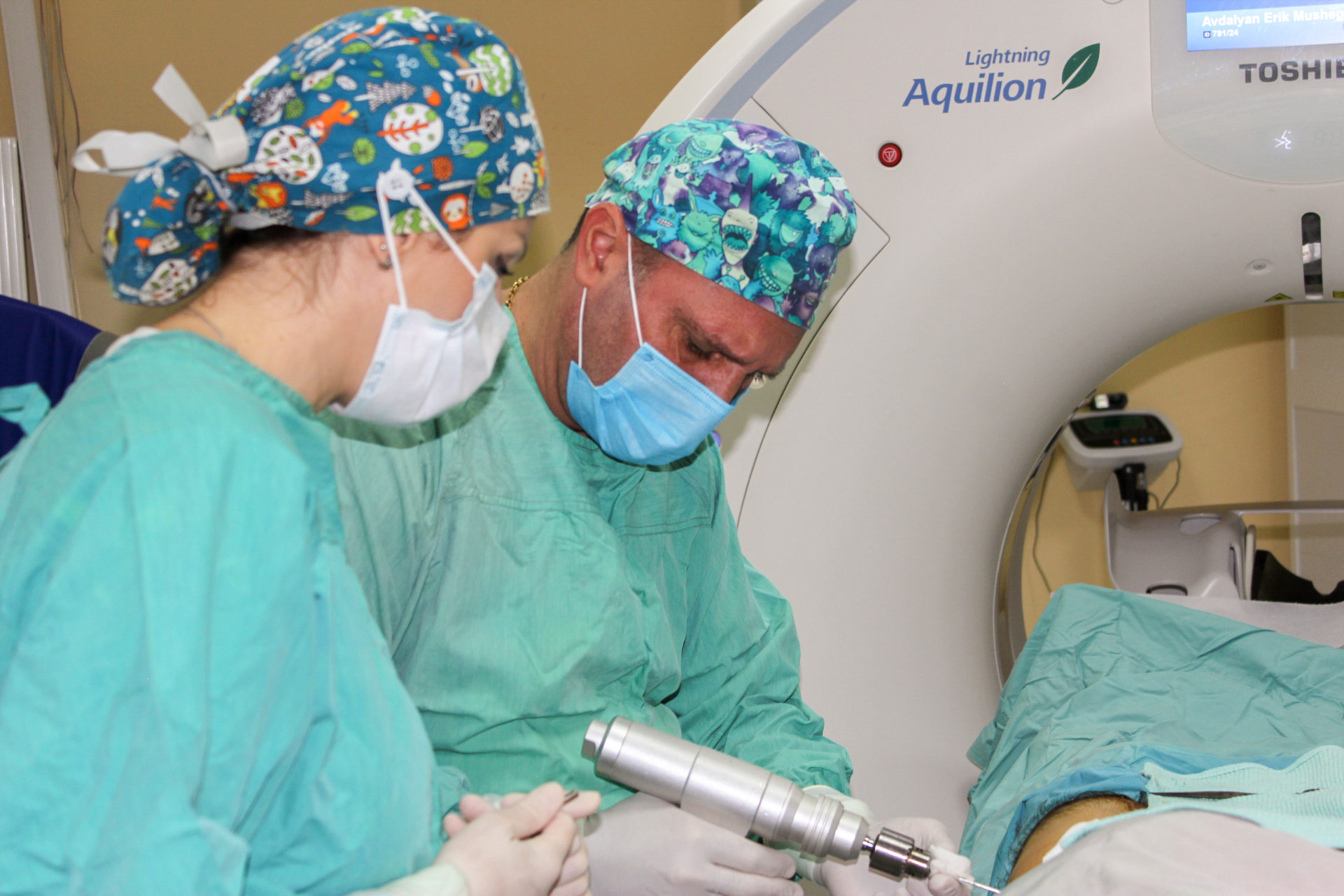
Another rare case, another team victory at the "Arabkir" Medical Center.
A 17-year-old patient had been complaining of pain in the left hip joint for three years and recently started limping. He had undergone several examinations and, as usual, consulted a rheumatologist, receiving treatment for hip osteoarthritis. However, as the treatment showed no effectiveness, the patient was referred to the "Arabkir" Medical Center for an orthopedic consultation. The orthopedic team suspected osteoid osteoma and ordered a computed tomography (CT) scan, which confirmed the presence of osteoid osteoma located in the neck of the femur.
Osteoid osteoma is a rare bone formation, occurring in 1-2 out of 100,000 people, most commonly in those aged 5-20. Its origin often remains unknown, and it can be located in various parts of the bone, more frequently in the distal parts of the tubular bones. One of the key symptoms is pain, especially at night.
To alleviate the pain significantly impacting the patient's quality of life, a surgical intervention was planned under computed tomography guidance. This technique allowed the surgical team to identify the shortest, least traumatic, and most effective path for surgical instruments to access the osteoma without damaging other important anatomical structures.
After carefully selecting the optimal access point, the orthopedic surgeon inserted a metal guide wire into the pathological focus, made a small 1 cm incision at the entry point, and completely excised the osteoma using a special scalpel. The entire procedure was monitored in real-time by a radiologist who reviewed the CT images.
The patient experienced immediate relief following the surgery, with complete resolution of the pain and restoration of a balanced gait.